Time’s Fun When You’re Having Flies

An introduction to my project

For this blog I will be talking about my research project this year. I’ve been studying the novel ribosomal regulatory mechanism of a hox gene discovered by Professor Andrew Renault (my supervisor). The Eukaryotic ribosome consists of 79-80 proteins which form a piece of machinery capable of interpreting genes to create amino acid sequences and proteins in  a process called translation. This works by reading the sequence of  a molecule called mRNA, a copy of the gene encoded in the genome. While this is the main role of the ribosome, there is mounting evidence of additional regulatory roles. Hox genes are a type of gene which control the body plan and patterning; they are characterised by the presence of a homeobox domain which is able to bind to specific sequences of DNA. Mutations that affect hox genes will therefore often cause changes to the body plan. One of the most well-known examples of this is the experiment by Malicki, Schughart, and McGinnis (1990) which used a mouse hox gene to make legs grow out of a  fly’s head instead of antenna.


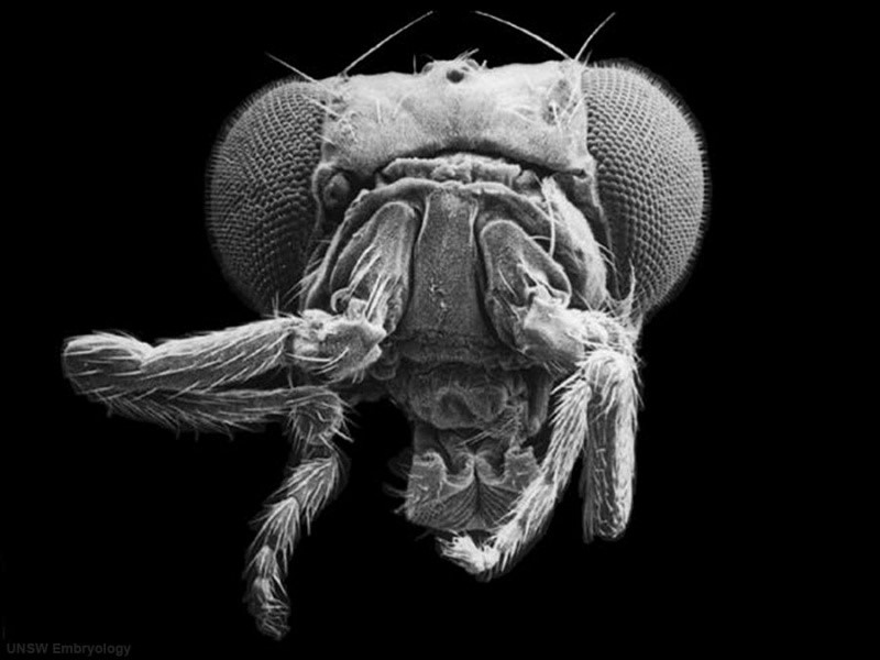
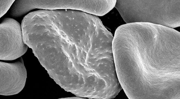
Fig.1 – Drosophila Melanogaster Mutant Antennapedia Head scanning EM (UNSW Embryology)

The ribosomal protein that I am investigating is called RPL39, a protein located at the exit tunnel of the ribosome where the amino acid chain emerges. Petrone et al. (2008) predicted that it may interact with another part of the ribosome called the 23S-rRNA tetraloop, working together to obstruct the tunnel exit.

Fig.2 – Diagram of a ribosome during translation, and approximate location of RpL39 (Adapted from
Renault publications)

There is another gene called Xrp1 which has been indicated to be highly involved in the phenotypes of many Ribosomal protein (Rp) mutations. Lee et al. (2018) found elevated Xrp1 in many Rp mutations, and Xrp1 has been linked to the minute phenotype. The minute phenotype is seen in 66/79 Rp mutants, and is characterised by shorter, finer bristles, particularly on the head, thorax, and scutellum (fig.3), an extended developmental period, lowered fertility, and reduced viability. It was shown that it is possible to save Rp mutant flies from this delay by introducing an Xrp1 mutation. This suggests that Rp mutations are unlikely to directly cause growth delay, rather a different pathway involving Xrp1 is affected.

Fig.3 – Pseudocoloured Bristles Displaying the Minute phenotype
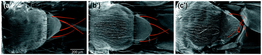
(a’) Wild type. (b’) RpS131 heterozygotes (minute). (c’) RpL141 heterozygotes (minute). (Marygold et. al 2007)

The discovery of a new mechanism

A mutant was discovered which caused the gonads to fail to coalesce; this mutation (A44) was affecting a well conserved proline amino acid to a serine.

Fig.4 – Alignment displaying the Conservation of Proline in RpL39 (Renault publications)

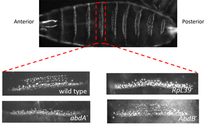
Such a change to body plan would not normally be anticipated from a ribosomal mutation, so more research was conducted, and it was discovered that the abnormal abdominal band pattern of A44 flies was identical to that of flies with a mutation in the hox gene abdominal-A (AbdA).

Fig.5 – Comparison of Abdominal Denticle Bands
(Renault publications)

When the protein abundance of AbdA and Tubulin was measured in A44 mutants and wild type flies it was shown that AbdA protein expresion was significantly reduced. Tubulin is a very prevalent protein found in all cells and could be used to represent general translation. Figure 6 suggests that RpL39 specifically affects AbdA expression not global expression. It was found that the mRNA remained unaffected, showing regulation of the protein either during or after translation.

Fig.6 – A Western Blot Showing a Decrease of AbdA Translation in A44 mutants
(Renault publications)

It was then found that changing the 5’ and 3’ untranslated regions (the parts of the mRNA sequence that aren’t translated) had no effect on the protein expression, meaning the sequence of the coding sequence must be causing the change. Preliminary experiments have shown that the N-terminus (the end of the protein sequence) is essential for this misfolding.

So Where Do I Fit Into All This?

This is where I come in: I’m working on several experiments to contribute to the paper being published on this mechanism.

Interaction study

I’m doing interaction studies by crossing virgin female A44 homozygous mutants and wild type flies with various heterozygous mutant males. I then wait for the flies to hatch and score the number of flies displaying the paternal mutation using various phenotypic markers. For example, I conducted a cross using RpS13 heterozygotes, using a marker called CyO to identify the non-mutants which will have curled wings. Once I have all the phenotypes talied up I will compare the ratio to the expected 50:50 Mendelian ratio. A reduced ratio of RpL39 RpS13 flies to the control RpS13 flies would indicate an interaction of some kind.

Fig.7 – CyO Phenotype
A: Wild Type B:CyO (curly wing) Phenotype

XRP1 localisation

To visualise the spatial distribution of of XRP1 in A44 embryos, I have made RNA probes for the final exon of XRP1 which I will be using for a colourmetric insitu hybridisation. To create these probes I used in vitro transcription on a plasmid, which I made via TA cloning an Xrp1 sequence into the PCR II TOPO vector. These probes will be used to stain fixed embryos, binding to the mRNA displaying the localisation of XRP1 which I will be able to view via a microscope.  Increased expression of XRP1 in A44 mutants would indicate that XRP1 is responsible for some of the minute phenotypic characteristics.

RPL39 and developmental timing

To confirm a delayed developmental timing I am setting up bottles of flies containing wild type, A44, and RpS13 (which displayes a minute phenotype) impregnated females. I will track the emergence of the different genotypes based on phenotypic markers.

Effect of AbdA N-terminus on GFP expression

I am also investigating if the N-terminus of AbdA alone is enough to cause GFP to misfold. This will be achieved by crossing RpL39 Nullo-Gal4 virgin females with GFP- AbdA N-terminus males. Nullo-Gal4 is a transcription factor required to trigger the expression of the GFP construct, allowing it to be seen in the embryos. These embryos will be killed and frozen for use in western blots to determine the protein levels.

My experience of this project

One of the greatest parts of doing this project has been seeing the inside of a scientific discovery. This mechanism has never been seen before, and it is amazing to be involved in investigating it. The hardest thing about my project so far has been the difficulty  of setting up crosses. This is due to the misformed gonads seriously impeeding fertility which has led to many of my crosses failing to successfully take. However, I must thank these malformed gonads as they helped direct the discovery of this facinating new mechanism.

This kind of research is important, furthering our understanding of gene regulation mechanisms creates the foundations for therapies to treat genetic disease. There are many diseases (including many cancers) asociated with ribosomal mutations, termed ribosomopathies, and there is often not a great deal of understanding of how the disease is caused. It is becoming increasingly more apparent that Rp’s have unique roles other than just contributing to translation. It may seem odd to some to investigate fly mutations to learn about ourselves, but 75% of all human diseases have Drosophila homologues. Their short generation time, size, and low cost, along with the bounty of fly genetic resources have made them perfect for investigating such research. While they say time flies when you’re having fun, it appears the reverse is also true.

Bibliography

Bastide, A. and David, A. (2018) ‘The ribosome, (slow) beating heart of cancer (stem) cell’, Oncogenesis. Nature Publishing Group, 7(4), p. 34. doi: 10.1038/s41389-018-0044-8.

Lee, C.-H. et al. (2018) ‘A Regulatory Response to Ribosomal Protein Mutations Controls Translation, Growth, and Cell Competition’, Developmental Cell. Cell Press, 46(4), p. 456–469.e4. doi: 10.1016/J.DEVCEL.2018.07.003.

Marygold, S. J. et al. (2007) ‘The ribosomal protein genes and Minute loci of Drosophila melanogaster’, Genome Biology. BioMed Central, 8(10), p. R216. doi: 10.1186/gb-2007-8-10-r216.

Petrone, P. M. et al. (2008) ‘Side-chain recognition and gating in the ribosome exit tunnel.’, Proceedings of the National Academy of Sciences of the United States of America. National Academy of Sciences, 105(43), pp. 16549–54. doi: 10.1073/pnas.0801795105.

Xue, S. and Barna, M. (2012) ‘Specialized ribosomes: a new frontier in gene regulation and organismal biology’, Nature Reviews Molecular Cell Biology. Nature Publishing Group, 13(6), pp. 355–369. doi: 10.1038/nrm3359.

Enlisting Symbiotic Bacteria to Help Fight Malaria at its Source

A summary of the paper “Driving mosquito refractoriness to Plasmodium falciparum with engineered symbiotic bacteria”

This week I have been investigating an inventive solution to one of the world’s greatest problems. I’ve long been interested in Malaria and its treatment, in part due to inspiration from my older brother working at the London School of Hygiene & Tropical Medicine. Malaria is a chronic infection caused when animals are bitten by mosquitos carrying Plasmodium parasites. According to the 2018 World Malaria Report the progress that had previously been made to reduce the severe prevalence of Malaria had stalled, with an estimation of 219 million cases and 435 thousand related deaths in 2017. Malaria is a disease caused by infection of the Plasmodium parasite spread by the Anopheles mosquito. The typical malaria managements are anti-parasitic drugs and vector control, but these methods are proving inadequate to stem the tide. This incredible problem is going to require some creative solutions.

For this blog I am summarising the work of Wang et al. 2017, a fascinating paper which opened up some incredible concepts for me. Sibao Wang and his team are testing paratransgenesis, a technique using genetically engineered symbiotic bacteria to prevent the malaria from infecting mosquitos (refractory) before they can infect humans, breaking the cycle. The most effective way to attack the plasmodium is at the Oocyst stage in the mosquito gut as there is likely to be only 1-5 that will survive the gut conditions before proliferating back out to thousands within the mosquito and then millions within humans.


Fig. 2. The malaria parasite cycle in the mosquito vector: (a) Life cycle of Plasmodium in the mosquito. The approximate developmental time at which each stage occurs in Plasmodium berghei (maintained at 20°C) is indicated. (b) Plasmodium parasite numbers undergo a severe bottleneck during its development in the mosquito gut. (Wang and Jacobs-Lorena, 2013)

This technique works by using a gut bacteria already found in mosquitos and inserting genes into them which confer malarial resistance to the mosquito. Wang et al. (2017) used the bacterial strain Serratia AS1 as it is also able to colonise the reproductive systems and haemolymph allowing for transition via sex and birth, proliferating the bacteria through a population. An important characteristic for paratransgenesis is for the symbiotic bacteria to not affect the fitness of the host. To test this Wang and his team investigated the fertility, fecundity, life span and blood feeding behaviour and found no obvious negative effects. Wang also showed that this strain would proliferate after blood meal, when the plasmodium would be introduced to the gut.

It is easy to forget that mosquitos don’t like malaria either as hosting a parasite decreases host fitness and due to this, it has been found that refractory mosquitos have increased fitness compared to their infected counterparts when feeding on malaria infected blood (Marrelli et al. 2007). This advantage is an important aspect of paratransgenesis as it will help prevent the mosquitos from developing immunity against the bacteria. The ideal bacteria would be an obligate symbiont, which is required for the mosquito to survive, however, one has not been discovered yet, if they even exist. I wonder if this increased fitness due to paratransgenesis could increase the mosquito population, increasing the prevalence of other mosquito diseases such as the dengue virus.

Fig. 2. Serratia AS1 bacteria stably colonize the midgut and rapidly proliferate after a blood meal. Bacteria colony forming units (CFUs) determined by measuring fluorescence of eGFP tagged bacteria

Wang introduced male and female mosquitos infected with AS1 (expressing GFP and mCherry respectively) as 5% of a 400 mosquito population. Of the next three generations, every member displayed both the paternal and maternal infection.

Fig. 3. Serratia AS1 bacteria efficiently spread throughout multiple mosquito generations. CFUs per fourth-instar larva gut.

Wang used the fusion protein Multi, made of 5 antimalarial peptides: MP2 (mutated bee venom phospholipase A2), scorpine, EPIP (enolase-plasminogen interaction peptide), Shiva1, and SM1 (salivary gland and midgut binding peptide 1). One way Plasmodium invades through the gut wall is by using a ligand to bind to a gut wall receptor and trick the gut into letting them through, like using a key to open a backdoor out of their gut. EPIP, MP2 and SM1 all prevent invasion by blocking parts of this system, EPIP binds to a Plasmodium ligand and MP2 and SM1 bind to mosquito endothelium (gut wall) receptors, while Scorpine and Shiva1 directly kill the parasites. To increase the potency of Multi the E. coli haemolysin A secretion system was used to export it into the midgut where the ookinetes will be. Multiple antimalarial peptides are required to help prevent drug resistance and increase efficacy. To test them in vivo AS1 engineered to produce the various peptides were fed on mosquitos via sugar meals 48 hours before a feeding on P. falciparum infected blood meal. Multi and scorpine were shown to be the most effective reducing oocyst loads by 92 and 93% respectively (figure 4).

Fig. 4. Inhibition of P. falciparum infection of A. gambiae by recombinant Serratia strains that are engineered to secrete anti-Plasmodium effector molecules. circles represent the number of oocysts in individual midguts, and horizontal lines indicate the median number of oocysts per midgut.

Paratransgenesis is often compared with sterility gene drives, another technology making great strides in recent years for managing mosquito populations. A gene drive is a form of genetic engineering which is able to propagate through a population faster than traditional genes. However, this would not be a perfect method for malaria management as destroying large populations of mosquitos could have unpredicted effects on the ecosystem. Only a small handful of the 30-40 anopheles species are able to be genetically engineered, which means not all mosquitos can be managed this way. One of the biggest hurdles affecting the effectiveness of this technology is the sexual isolation between populations, preventing the gene flow required, meaning you could potentially wipe out one species in an area for another to immediately take its place. In contrast, all mosquito species contain symbiotic bacteria able to be modified, and many can be infected by the same ones, allowing a strategy impact across species.

One of the most attractive features of paratransgenesis is the low cost as culturing and transporting bacteria is cheap once the initial strains are engineered. Mancini et al. (2016) previously showed in a semi-field study that all you have to do to infect a population is provide sugar water containing the bacteria for the mosquitos to feed on.

While it’s obvious this technique is effective in a lab environment, there has been no current true field studies of yet. This is in part due to concerns people have about releasing genetically engineered organisms out into the world, largely as once released there is no way to recall them. However, such studies are not without precedent, the first field test of a genetically engineered microbe (GEM) was carried out back in 1987 releasing anti-frost bacteria across strawberry fields. These tests showed the bacteria to be safe and effective, but due to a long evaluation by the EPA and NIH alongside difficulties in gaining regulatory approval the research had become too costly and was dropped, discouraging future GEM research.

It is clear to me that paratransgenesis could be a useful weapon in the fight against malaria, but shouldn’t be done alone. The only way to defeat such an enemy is by fighting it on all fronts, antimalarial drugs, mosquito nets, insecticides, genetic techniques such as paratransgenesis and gene drives, and potentially one day vaccines will all likely play a large role.

Bibliography

Lindemann, J. & Suslow, T. V. Ecology and Epidemiology Competition Between Ice Nucleation-Active Wild Type and Ice Nucleation-Deficient Deletion Mutant Strains of Pseudomonas syringae and P. fluorescens Biovar I and Biological Control of Frost Injury on Strawberry Blossoms.

Mancini, M. V. et al. Paratransgenesis to control malaria vectors: a semi-field pilot study. Parasit. Vectors 9, 140 (2016).

Marrelli, M. T., Li, C., Rasgon, J. L. & Jacobs-Lorena, M. Transgenic malaria-resistant mosquitoes have a fitness advantage when feeding on Plasmodium-infected blood. 104, (PNAS, 2007).

Wang, S. & Jacobs-Lorena, M. Genetic approaches to interfere with malaria transmission by vector mosquitoes. Trends Biotechnol. 31, 185–193 (2013).

Wang, S. et al. Driving mosquito refractoriness to Plasmodium falciparum with engineered symbiotic bacteria. Science 357, 1399–1402 (2017).

World Health Organization. WORLD MALARIA REPORT 2018 ISBN 978 92 4 156565 3. (2018).

Cold Fusion – Fiction or Future?

What is Cold Fusion?

For this week I’ve been looking into cold fusion and the scandal of Fleishman and Pons’ work. So, to start things off, what is fusion? Fusion is a nuclear process where two atoms are fused to form one, compared with the current nuclear fission power where one atoms is split into two. For fusion to occur strong nuclear attraction must overcome electric repulsion, in effect this means there’s a barrier the atoms must push past to fuse. Deuterium, an isotope of hydrogen is normally investigated for fusion as helium requires a neutron to form, additionally hydrogen has the fewest electrons and so the lowest electric repulsion to overcome.

One of the biggest differences between these fusion methods is that fusion is able to generate far more energy while being environmentally cleaner, as deuterium is readily available in sea water and the only byproduct is helium. Additionally, unlike fission chain reactions, fusion is a stable reaction and so the possibility of a Chernobyl like event is reduced.

So now we know what fusion is and why people love it, but what about cold fusion? Well getting atoms close enough for the strong nuclear force to trigger fusion generally requires an awful lot of pressure and heat, generally around 100 million degrees. Current prototype fusion generators must be large to allow these conditions to be obtained, for example; pictured below is the International Thermonuclear Experimental Reactor currently under construction. Cold fusion is the concept of generating fusion energy at considerably lower temperatures, this could potentially allow the technology to be far smaller and the reaction be both easier and cheaper to maintain.

One of the most surprising things I discovered researching this topic was that cold fusion had actually been proven possible by Alverez et al. (1957). This was achieved by replacing hydrogen electrons with heavier muons, lowering their orbit decreasing the size of the atom (pictured below). This size changed allows the nuclei to get close enough for fusion to occur at room temperature. Muons are capable of catalysing multiple fusion reactions but their half-life is only 2.2 µs meaning each muon is only able to generate 2.7GeV, while requiring approximately 5GeV to create. Therefore, this method of fusion is unable to be adapted for generating electricity.

Enter Fleishmann and Pons

During the late 1960s professor Martin Fleischmann (who would go on to win the 1970 medal for electrochemistry and thermodynamics from the Royal Society of London) conducted experiments using palladium to separate hydrogen from deuterium. He found that due to a chemical reaction on its surface, palladium is able to absorb 900 times its own volume of hydrogen. During a conversation Fleischmann had with his prior mentee Stanley Pons while working at the University of Utah, the two realised that palladiums properties may force hydrogen atoms close enough to trigger fusion. This led them to design their fusion cell (pictured bellow) which used electrolysis to split deuterium water using a palladium electrode to absorb deuterium.

During experimental testing the cell temperature was measured at 100 times as would be expected without fusion, and neutrons were detected at levels indicating fusion. Excited Fleischmann and Pons rushed to publish, however, 18 months before their self set deadline another scientist, Steven Jones contacted them. Jones told them that he had been working on the same phenomenon but wasn’t looking at cold fusion as a power source and was ready to publish. Jones suggested a mutually beneficial collaboration as Jones was a nuclear physicist and could have great insight. However, Fleischmann and Pons convinced that Jones had used details from their grant application without permission, were unwilling to collaborate but did agree to publish simultaneously. Fleischmann and Pons however, didn’t keep their word and submitted 13 days ahead of the agreed date and announced during a press conference before the paper could be published, that they had created a sustained nuclear fusion reaction at room temperature causing a media explosion.

After the announcement many scientists were anxious to read the paper to see the proof of their high claims. To accommodate this, an abbreviated form of peer review was used. Once the paper was released many were quick to find huge flaws in their experiment including lacking controls and incorrect calculation of the force magnitudes within the palladium. The paper didn’t include all of their methodology and so others were unable to properly replicate the experiment for testing, this left people guessing resulting in a wide range of conflicting results, with most of the supporting papers being redacted due to errors.

An internal investigation showed that Fleischmann and Pons neutron data had likely been the result of equipment error, to account for this Pons claimed that it was possible the helium was being absorbed by the palladium before it could emit neutrons. However, when tested one of their rods didn’t display elevated levels of helium, in response to this Pons admitted that the rod in question hadn’t produced as much heat as he initially claimed.

To attempt to validate the research the University of Utah had a fellow professor, Michael Salamon who using their lab and equipment was unable to detect elevated neutron levels. However, Pons claimed that these results were meaningless as Salamon hadn’t achieved fusion within the cells, indicated by a lack of increased temperature.

By the time a year had passed, due to the lack of both reproducibility and proof of fusion the scientific community as a whole deemed that the results were due to experimental error. By this point over 100 million dollars of tax payer money had been spent investigating their research, and the public nature of the debacle seriously affected the perception of science.

An end of cold fusion research?

Cold Fusion research does still occur but the scientists involved struggle for funding and high impact publication. In 2003 the United States Department of Energy conducted a review of cold fusion research. Approximately half of the reviewers deemed the experiments were producing heat with most agreeing inadequate adequate evidence of fusion. There are also still many prestigious scientists backing up cold fusion, such as Professor Brian D. Josephson of Cambridge, awarded a Nobel Prize in physics in 1973.

While it’s impossible for me to truly conclude if cold fusion power is possible, I do think Fleischmann and Pons believed they had achieved it, and Fleischmann maintained this claim until the day he died in 2012. I believe that much of their conduct was due to pressure placed on them by the University and their protective nature of their research. While Fleischmann and Pons may not have been purposefully false with their research their failure to conduct good, scientific and moral practise has led to the shunning of their field in science. To me, this is a perfect case study for how not to act when dealing with controversial science, research must be well structured, proper peer review must be followed, total honesty is always required, and collaboration is often key.

Bibliography

Alvarez, L. W., Bradner, H., Crawford, F. S., Crawford, J. A. 1957. Phys. Rev., 105: 1127

Ball, P. Martin Fleischmann (1927–2012). Nature 489, 34–34 (2012).

Brumfiel, G. US review rekindles cold fusion debate. news@nature (2004). doi:10.1038/news041129-11

Fleischmann, M., and S. Pons. 1989. Electrochemically induced nuclear fusion of deuterium. Journal of Electroanalytical Chemistry 261:301-308.

Jones, S.E., E.P. Palmer, J.B. Czirr, D.L. Decker, G.L. 1989. Observation of cold nuclear fusion in condensed matter. Nature 388:737-740.

Josephson, B. D. Cold fusion: Fleischmann denied due credit. Nature 490, 37–37 (2012).

Cold fusion: A case study for scientific behavior.